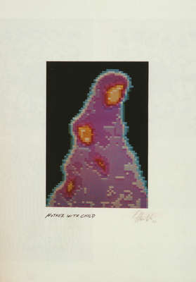
Scintigraphs/scintigrams were actually used for nuclear medical examinations and the depiction of inner organs, e.g. in tumour diagnosis. After the administration of marked radioactive substances that accumulate in the tissues to be examined, gamma rays are emitted. These are then recorded with the aid of a scanner or a gamma camera and transformed into light images. This creates scintigrams/line-raster images, in which the density of the lines indicates the distribution of the gamma rays and their activity.
These images may also be in colour, whereby different colours stand for different activity levels, e.g. red for a lot of activity. Artists such as Havlik and Herbert W. Franke used this principle to produce graphic works.
5


